The extraoral periapical radiographic technique was performed for both maxillary and mandibular teeth using Newman and Friedman technique2. Machine learning techniques th e more images in the dataset we.

Periapical Radiography Pocket Dentistry
Ensure they are seated high enough so it is easy to see the occlusal.
. The Long Cone Paralleling Technique. Students learn how to expose dental periapical x-ray film using the Rinn Paralleling Technique using the XCP film holder. 3 What is an intraoral technique of exposing periapical images.
In this video let see about the principle advantage and disadvantage of. The X-ray tubehead is then aimed at right angles vertically and horizontally to both the tooth and the image. By using a film sensor holder with still.
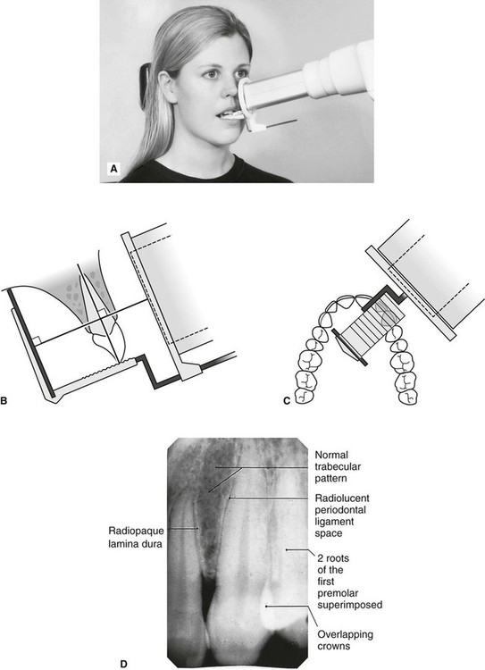
The X-ray is taken and the exposed plate is then loaded into a scanner or processor which reads the image. Periapical X-rays are used to detect any abnormalities of the root structure and surrounding bone structure. The bisecting-the-angle technique and the more commonly used long cone paralleling technique.
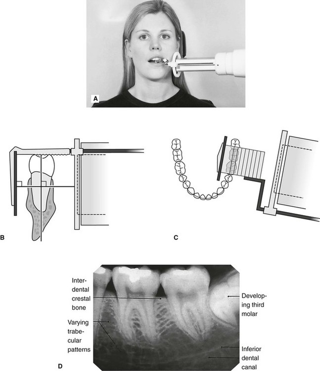
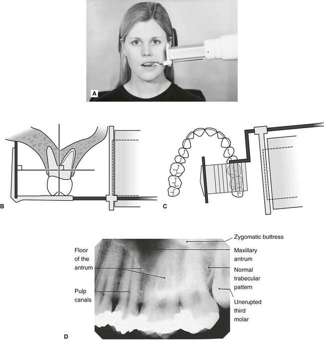
The film is placed parallel to the long axis of the tooth in question and the central x-ray beam should be directed perpendicular to the long axis of the tooth. Click to see full answer. Paralleling and Bisecting angle are the techniques used for periapical radiography.
The film is placed parallel to the long axis of the tooth in question and the central x-ray beam should be directed perpendicular to the long axis of the tooth. Each periapical X-ray shows all teeth in one portion of either the upper or lower jaw. Periapical images have been collected using the FONA X70 Intraoral X-rays machine and PSPIX Imaging Plates.
Periapical radiographic techniques Periapical radiography is designed to give diagnostic images of the apical portions of teeth and their adjacent tissues. Students are given a demonstration of panoramic radiology. With this technique the film is placed parallel to the long axis of a tooth allowing the X-ray to be focused perpendicular to the long axis of the tooth.
6 Which exposure technique does the American Academy of Oral and Maxillofacial Radiology and the American Dental Education Association recommend. Occlusal X-rays show full tooth development and placement 9. 4 Which technique is most often used when exposing a periapical image.
Periapical X-rays. Periapical X-rays detect any unusual changes in the root and surrounding bone structures. Intraoral periapical radiographs can be produced using two different techniques.
Inclusion criteria included periapical X-ray images of permanents teeth and patients aged 14 years old with good sharpness. Paralleling technique Bisecting angle technique Paralleling technique It is also called the extension cone paralleling technique right angle technique and long cone technique. Parallel technique The image receptor is placed in a holder and placed in the mouth parallel to the longitudinal axis of the tooth under.
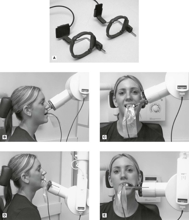
The patient was positioned upright with hisher mouth was opened as wide as possible to allow the X-ray beam to pass to the sensor unobstructed from the opposite side of the mouth. The paralleling technique results in good quality x-rays with a minimum of distortion and is the most reliable technique for taking periapical x-rays. The sensor was placed on the.
5 What is bisecting angle technique in dentistry. With these advances the need for more precise diagnostic tools specially imaging methods have become mandatory. A periapical x-ray or PA film will show one or two teeth in their entirety in one single image right from the crown of the tooth which is the part exposed in the mouth to the very tips of the tooth roots located in the jawbone as well as.
50 patients had their periapical dental radiographs taken utilizing the long cone paralleling technique. Extraoral radiograph Panoramic X-ray Tomograms Cephalometric projections Sialography Computed tomography 10. The paralleling technique results in good quality x-rays with a minimum of distortion and is the most reliable technique for taking periapical x-rays.
Paralleling Technique for Periapical X-rays The paralleling technique results in good quality x-rays with a minimum of distortion and is the most reliable technique for taking periapical x-rays. The film is placed parallel to the long axis of the tooth in question and the central x-ray beam should be directed perpendicular to the long axis of the tooth. Periapical X-ray images expor ting results and reading results.
The X-ray head is directed at right angles vertically and horizontally of both the tooth and the image receptor. The patient is seated upright in the dental chair and should remove any removable dental appliances glasses or jewelry that could interfere with the X-ray beam. A full mouth intraoral examination consists of 14 periapical radiographs with two bite-wing films and provides an image of all teeth and related structures.
Exclusion criteria were periapical X-ray images of tooth germs or images which have distortion effects. Basically there are two techniques for taking periapical radiography. Film development and mounting are discussed and practiced.
All radiographs were obtained by. Most frequently used radiography is for the periapical which is performed by the bisecting Thus when considering the execution of the radiographic technique and the possibility of errors that occur during the exposure of X-ray image XR receptors it is important to identify those that occur more frequently. From the simple intra-oral periapical X-rays advanced imaging techniques like computed tomography cone beam computed tomography magnetic resonance imaging and ultrasound have also found place in modern dentistry.
To take a periapical exposure the hygienist or x-ray technician places a small photosensitive imaging plate coated with phosphorus into a sterile wrapper and inserts it into the patients mouth just like a conventional X-ray film card. What are periapical radiographs used for. Have the more e ectively the m odel works.
Periapical X-rays are used to detect any abnormalities of the root structure and surrounding bone structure. By using a filmsensor holder with fixed image receptor and. The image receptor is placed in a holder and positioned in the mouth parallel to the long axis of the tooth under.

How Make Periapical X Ray
Periapical Radiography Clinical Gate

How Make Periapical X Ray

Periapical Radiography Pocket Dentistry
Periapical Radiography Clinical Gate
Periapical Radiography Clinical Gate
Periapical Radiography Clinical Gate
Periapical Radiography Pocket Dentistry
0 comments
Post a Comment